Different Methods of Diagnosis
Typical diagnosis occurs when a patient is experiencing the three prominent signs that are most evident in Parkinson’s disease: resting tremor, rigidity, and bradykinesia. Once a patient is diagnosed and is put on the medication Levodopa, if the individual does not respond to the medication but continues to show signs and symptoms of Parkinson’s disease this is referred to as Idiopathic (unknown) Parkinson’s Disease.
New methods of MRI scanning are being used to detect early signs of PD
“Conventional MRI cannot detect early signs of Parkinson's, so the Oxford researchers used an MRI technique, called resting-state fMRI, in which people are simply required to stay still in the scanner. They used the MRI data to look at the 'connectivity', or strength of brain networks, in the basal ganglia – part of the brain known to be involved in Parkinson's disease.” (University of Oxford, June 2014).
By defining a threshold level of connectivity, researchers were able to detect PD patients with 100% accuracy and non PD patients with 89% accuracy(few false positives). By testing the strength of the brain networks in the basal ganglia, diagnosis of Parkinson’s can begin at even the earliest stages of the disease. The research team hopes to take this to the next step of picking up PD before symptoms arise in the future.
Alpha-Synuclein Protein
“Alpha-synuclein is the protein, component of the lewy bodies and its accumulation in tissues helps diagnose PD in patients. Abnormal alpha-synuclein aggregation may begin in the peripheral nervous system, possibly in the nerves of the gastrointestinal submucosal region many years before motor symptoms appear. In one study, colon tissue extracted during a colonoscopy was analyzed in patients in the early stages of PD but who had not been treated for PD. Tissue samples showed that 9 out of 10 had alpha-synuclein inclusions in the tissue” (Christine, 2011a).
Screening for Biomarkers
Biomarkers is playing a very important role in diagnosing specific diseases and supporting to diagnose Parkinson’s disease to a very limited extent. A new developed technique called diffusion MRI have shown great improvement in detecting its signs and also to discriminate between the .typical and atypical type of Parkinson’s disease.
“The Michael J. Fox Foundation is using these techniques in an ongoing study of biomarkers called the Parkinson’s Progression Markers Initiative (PPMI). They are looking at movement, cognitive, and brain biomarkers in addition to blood, urine, DNA, and spinal fluid sampling in 400 newly diagnosed PD patients over a 3- to 5-year period (Christine, 2011a)”.
Mathematical Models for Dopaminergic and Serotonergic Systems
J.A. Best and his team have created mathematical models for the serotonin and dopamine terminals. They used blood tryptophan as an input in the model and other nine substrates can also be used as an input in the model. For each differential equation that has been used to quantitate mass balance expression, “The rate of change of the concentration of a substrate is simply the sum of the rates of the reactions by which it is made minus the sum of the rates of the reactions in which it is used. For example, the concentration of 5-hydroxytryptophan, [5htp], satisfies:
J.A. Best and his team have created mathematical models for the serotonin and dopamine terminals. They used blood tryptophan as an input in the model and other nine substrates can also be used as an input in the model. For each differential equation that has been used to quantitate mass balance expression, “The rate of change of the concentration of a substrate is simply the sum of the rates of the reactions by which it is made minus the sum of the rates of the reactions in which it is used. For example, the concentration of 5-hydroxytryptophan, [5htp], satisfies:
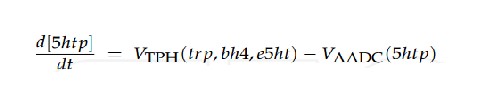
VTPH is the velocity of the TPH reaction and VAADC is the velocity of the AADC reaction. One must specify exactly how these velocities depend on the current values of various substrates. VTPH is given by:
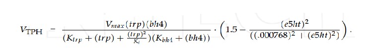
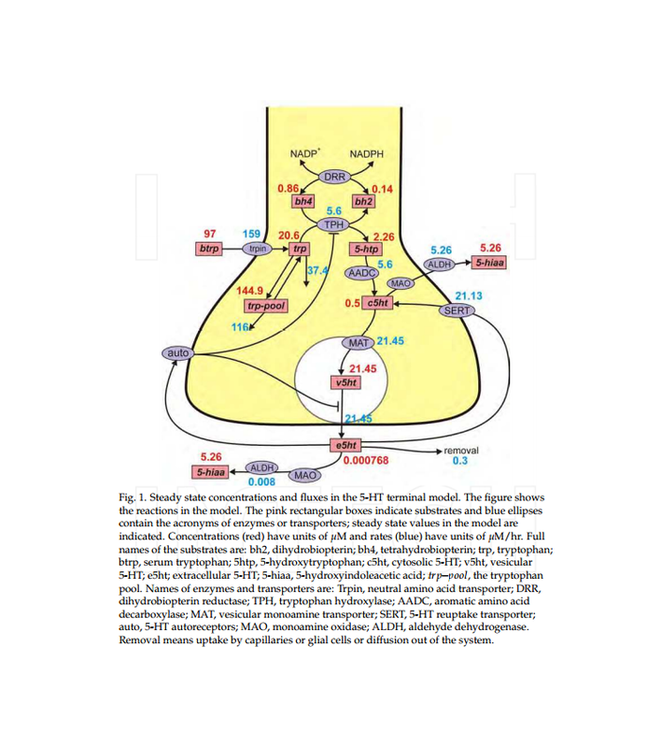
The first term on the right is of Michaelis-Menten form and gives the dependence of the velocity on the concentrations of tryptophan and bh4. The enzyme TPH shows substrate inhibition (Best et al., 2010a; Friedman et al., 1974; McKinney et al., 2005), which is the reason for the (trp) 2 term in the denominator. The second term on the right expresses how the concentration of extracellular 5-HT influences the rate of synthesis via the auto receptors. At normal e5-HT concentration (.768 nM) this factor equals one. As e5-HT goes up the factor can go as low as 0.5 and as e5-HT goes down, the factor can go as high as 1.5. We chose Km and Ki values from the literature and chose the Vmax so that the normal velocity of the the TPH reaction is in the range given by experiments. The form of the second factor is more speculative. Though it is certain that increasing extracellular concentrations of 5-HT inhibit synthesis via the auto receptors (Adell et al., 2002), there is relatively little information in the literature about the range of e5-HT concentrations over which the effect takes place and about the strength of the effect in the low nanomolar range.”(Best et.al, 2009,2010a,2010b).
Titcombe et.al states that, "There are lot of questions which have been unanswered regarding Parkinson’s disease. Recent studies have found two hypothesis for the question: 'Where does the tremor in Parkinson’s disease originate from'? Researchers stated that this process mainly involves central and peripheral feedback mechanisms. They stated that oscillations originate from central nervous system may be somewhere in motor control regions of the brain such as thalamus and basal ganglia."
Mathematical Model for Parkinson’s Tremor
“In the 1960’s, two articles appeared (Austin and Tsai 1962, Austin et.al; 1965) describing a model for Parkinson’s tremor based on the Van der Pol oscillator. The Van der Pol equation is of the form:

...This equation is second-order, nonlinear, and depends on one parameter, α.”(1965,1962).
Coping
We must remember that this disease is more than formulas and equations, this is people's lives. If you or a loved one is suffering from Parkinson's Disease the big idea from this site we hope you can take to heart is that, you are not alone.
We must remember that this disease is more than formulas and equations, this is people's lives. If you or a loved one is suffering from Parkinson's Disease the big idea from this site we hope you can take to heart is that, you are not alone.
Researchers are learning more about this disease everyday, and as students we must remember that whatever specialty we choose to follow in the field of biomedical engineering, that the end goal is improving people's lives.
References
Articles/Websites:
1. Adell, A., Celada, P., Abella, M. T. & Artigasa, F. (2002). Origin and functional role of the extracellular serotonin in the midbrain raphe nuclei, Brain Res Rev 39: 154–180.
2. Aminoff MJ, Christine CW, Friedman JH, et al. (2011, March). Management of the hospitalized patient with Parkinson’s disease: Current state of the field and need for guidelines. Parkinsonism Relat Disord. 17(3): 139–45. Retrieved August 30, 2013 from <http://www.ncbi.nlm.nih.gov/pmc/articles/PMC3070297/?tool=pubmed>.
3. Aminoff MJ. (2010). Young-Onset Parkinson’s Conference (Video). 18 Aug 2013. <http://www.hindawi.com/journals/pd/2012/198316/cta/.ttp://pdcenter.neurology.ucsf.edu/videos/gene-therapy-pd>.
4. Barcia C. (2013). Glial-Mediated Inflammation Underlying Parkinsonism. Scientifica vol. 2013, Article ID 357805. Retrieved August 15, 2013 from <http://www.hindawi.com/journals/scientifica/2013/357805/>.
5. Best, J. A., Nijhout, H. F. & Reed, M. C. (2009). Homeostatic mechanisms in dopamine synthesis and release: a mathematical model, Theor Biol Med Model 6: 21.
6. Best, J. A., Nijhout, H. F. & Reed, M. C. (2010a). Models of dopaminergic and serotonergic signaling, Pharmacopsychiatry 43(Supp. 1): 561–566. Mathematical Models: Interactions Between Serotonin and Dopamine in Parkinson’s Disease 415. <www.intechopen.com>.
7. Best, J. A., Nijhout, H. F. & Reed, M. C. (2010b). Serotonin synthesis, release and reuptake in terminals: a mathematical model, Theor Biol Med Model 7: 34.
8. Bonnet AM, Jutras MF, Czernecki V, et al. (2012). Nonmotor symptoms in Parkinson’s disease in 2012: Relevant clinical aspects. Parkinson’s Disease vol. 2012, Article ID 198316. Retrieved January 31, 2013 from <http://www.hindawi.com/journals/pd/2012/198316/cta/>.
9. Bouwmans AEP, Vlaar AMM, Mess WH, Kessels A, and Weber WEJ. (2013). Specificity and sensitivity of transcranial sonography of the substantia nigra in the diagnosis of Parkinson’s disease: prospective cohort study in 196 patients. BMJ Open. 2013; 3(4): e002613. Retrieved August 17, 2013 from <http://bmjopen.bmj.com/content/3/4/e002613>.
10. Christine C. (2011a). Biomarkers for Parkinson’s Disease. (Video). 15 Aug. 2013. <http://pdcenter.neurology.ucsf.edu/videos/biomarkers-Parkinson-s-disease>.
11. Christine C. (2011b). Hospitalization of the Parkinson’s Patient (video). Parkinson’s Disease Clinic and Research Center. Retrieved August 29, 2013 from <http://pdcenter.neurology.ucsf.edu/videos/hospitalization-Parkinson-s-patient>.
12. "Environmental Factors and Parkinson's: What Have We Learned?" - Parkinson's Disease Foundation (PDF). Web.
13. Fakhar K, Hastings E, Butson CR, et al. (2013). Management of deep brain stimulator battery failure: Battery estimators, charge density, and importance of clinical symptoms. PLoS ONE 8(3): e58665. Retrieved August 21, 2013 from <http://www.plosone.org/article/info%3Adoi%2F10.137
1%2Fjournal.pone.0058665>.
14. Feher, Joseph J. Quantitative Human Physiology: An Introduction. Amsterdam: Elsevier/Academic, 2012. 352. Print.
15. Granado N, Ares-Santos S, Moratalla R. (2013). Methamphetamine and Parkinson’s disease. 17 June. 2013. <http://www.hindawi.com/journals/pd/2013/308052/>.
16. Hauser. “Parkinson’s Disease” medscape aug 15, 2014 <http://emedicine.medscape.com/article/1831191-overview#aw2aab6b2b4aa>.
17. Hohler AD, Tsao JM, Katz DI, et al. (2012). Effectiveness of an inpatient movement disorders program for patients with atypical parkinsonism. Parkinson’s Disease vol. 2012, Article ID 871974, 6 pp. Retrieved August 9, 2013 from <http://www.hindawi.com/journals/pd/2012/871974/>.
18. Imperical College.(2014).<http://www3.imperial.ac.uk/newsandeventspggrp/imperialcollege/newssummary/news_9-1-2014-16-19-43>.
19. Jensen P, Krabbe C, Meyer M. (2011). Cell therapy for Parkinson’s disease. In David Finkelstein (ed.), Status and Perspectives: Towards New Therapies for Parkinson’s Disease.ISBN: 978-953-307-463-4, InTech, DOI: 10.5772/21203. Retrieved October 11, 2013 from <http://www.intechopen.com/books/towards-new-therapies-for-Parkinson’s-disease/cell-therapy-for-Parkinson-s-disease-status-and-perspectives>.
20. Kwan LC, Whitehill TL. (2011). Perception of speech by Individuals with Parkinson’s disease: A review. Parkinson’s Disease vol. 2011, Article ID 389767, 11 pp. Retrieved April 22, 2013 from <http://www.hindawi.com/journals/pd/2011/389767/>.
21. Lescaudron, Laurent. Stem Cells and Neurodegenerative Diseases. CRC, 2014. Print.
22. “Medical Dictionary” free online medical dictionary. <http://medical-dictionary.thefreedictionary.com/>.
23. Models: Interactions Between Serotonin and Dopamine in Parkinson’s Disease. 415. <www.intechopen.com>.
24. "MRI Brain Scans Detect People with Early Parkinson's." University of Oxford. 12 June 2014. Web. <http://www.ox.ac.uk/news/2014-06-12-mri-brain-scans-detect-people-early-parkinsons>.
25. National Human Genome Research Institute (NHGRI). (2011). Learning About Parkinson’s Disease. Retrieved July 12, 2013 from <http://www.genome.gov/10001217#2>.
26. National Institutes of Health (NIH). (2013a). Genetics Home Reference. Retrieved August 8, 2013 from <http://ghr.nlm.nih.gov/condition/Parkinson-disease/show/Related+Gene(s)>.
27. "NINDS Deep Brain Stimulation for Parkinson's Disease Information Page." Deep Brain Stimulation for Parkinson's Disease Information Page: National Institute of Neurological Disorders and Stroke (NINDS). Web.
28. Okun MS. (2012). Too many pills: Improving delivery systems for Parkinson’s disease drugs. Retrieved October 10, 2013 from <http://Parkinson’s.org/Patients/Patients---On-The-Blog/May-2012/Too-Many-Pills--Improving-Delivery-Systems-for-Par>.
29. Okun MS, Hassan A. (2012). Hospitalization: An action plan to be aware in care. National Parkinson’s Foundation. Retrieved August 29, 2013 from <http://event.netbriefings.com/event/npf/Archives/hospitalization/>.
30. “Parkinson’s disease Information”. Parkinson.org updated on June 23, 2009. <http://www.parkinsons.org/index.html>.
31. Parkinson J. (1817). Essay on the Shaking Palsy. Retrieved January 16, 2013 from <http://books.google.com/books?id=4ygSAAAAYAAJ&printsec=frontcover&source=gbs_ge_summary_r&cad=0#v=onepage&q&f=false>.
32. Robertson, Lauren.,B.A, MPT. A Train Education, Continuing Education for Health Professionals. “Parkinson’s Disease: Moving Forward.” <https://www.atrainceu.com/course-module/1941275-085_parkinsons-module-15>.
33. “Statistics on Parkinson’s Disease” Parkinson’s Disease Foundation ©2014 Parkinson's Disease Foundation, Inc. <http://www.pdf.org/en/parkinson_statistics>.
34. Tambasco N, Simoni S, Marsili E, et al. (2012). Clinical aspects and management of levodopa-induced dyskinesia. Parkinson’s Disease vol. 2012, Article ID 745947. Retrieved February 5, 2013 from <http://www.hindawi.com/journals/pd/2012/745947/>.
35. Tarsy, Daniel. "Neuroprotective Therapy for Parkinson Disease." Neuroprotective Therapy for Parkinson Disease. Web.
36. "The Lancet IPad App: Articles in a New Light." Long-term Safety and Tolerability of ProSavin, a Lentiviral Vector-based Gene Therapy for Parkinson's Disease: A Dose Escalation, Open-label, Phase 1/2 Trial : The Lancet. Web.
37. "The Lancet IPad App: Articles in a New Light." Neural Transplantation for the Treatment of Parkinson's Disease : The Lancet Neurology. 1 July 2003. Web. <http://www.thelancet.com/journals/laneur/article/PIIS1474-4422(03)00442-3/fulltext>.
38. University of Luxembourg. "Transplanted induced neural stem cells become fully integrated in the neuronal network in mice."Medical News Today. MediLexicon, Intl., 7 Aug. 2014. Web. <http://www.medicalnewstoday.com/releases/280631.php>. 19 Nov. 2014.
39. Wikipedia contributors. "L-DOPA." Wikipedia, The Free Encyclopedia. Wikipedia, The Free Encyclopedia, 2 Nov. 2014. Web. 25 Nov. 2014.
40. Wikipedia contributors. "Parkinson's disease." Wikipedia, The Free Encyclopedia. Wikipedia, The Free Encyclopedia, 7 Nov. 2014. Web. 24 Nov. 2014.
Images:
1. Basal Ganglia Components. Digital image. Basal Ganglia. N.p., n.d. Web. 24 Nov. 2014. <www.mybrainfacts.com>.
2. Beautiful PD Tulip Pin. Digital image. Lasting Impressions. N.p., n.d. Web. 23 Nov. 2014. <lastingimpressionspins.com>.
3. Brain imaging showing the stages of Parkinson's disease. Digital image.Reuters. Reuters/NIH, 4 Apr. 2012. Web. 23 Nov. 2014. <www.reuters.com>.
4. Brain Regions Affected By Parkinson's Disease. Digital image. Fresh Jinks. N.p., n.d. Web. 23 Nov. 2014. <freshjinks.com>.
5. Deep Brain Stimulation. Digital image. Medscape. N.p.,n.d.Web. 23 Nov. 2014. http://img.medscape.com/fullsize/migrated/498/456/jnn498456.fig6.gif
6. Dopamine Levels in Parkinson’s Disease. Digital image. Health Resource 4 U. N.p., n.d. Web. 24 Nov. 2014. <www.healthresource4u.com>.
7. Drawing of a Lewy Body. Digital image. Parkinson's Disease: Frequently Asked Questions. NIH Senior Health, n.d. Web. 23 Nov. 2014. <nihseniorhealth.gov>.
8. Embryo Stem Cells. Digital image. N.p., n.d. Web. 23 Nov. 2014. http://www.impactaging.com/papers/v3/n5/full/100328/Figure1.jpg
9. Fluorodopa PET Scan. Digital image. A Train Education. N.p., n.d. Web. 23 Nov. 2014. <www.atrainceu.com>.
10. Gene Therapy. Digital image. N.p., n.d. Web. 23 Nov. 2014. http://www.engage-science.com/wp-content/uploads/2012/08/shi_julie_graphic.jpg
11. Great Scott! Could Time Travel Be a Movement Disorder? Digital image. The NEI Connection. NeuroRelief, 04 June 2013. Web. 24 Nov. 2014. <neuroendoimmune.wordpress.com>.
12. Hernandez, Brian. Dopamine Levels. Digital image. Brandeis University. Proteopedia, 29 Nov. 2011. Web. 23 Nov. 2014. <blogs.brandeis.edu>.
13. Lewy Body. Digital image. Department of Pathology, University of Oklahoma Health Sciences Center, n.d. Web. 23 Nov. 2014. <http://moon.ouhsc.edu/kfung/jty1/NeuroTest/Images/SampleR03-Lewybody.gif>.
14. Nervous System Diseases. Digital image. WebMD. Parkinson's Disease Health Center, n.d. Web. 23 Nov. 2014. <http://www.webmd.com/parkinsons-disease/>.
15.Obeso, Jose A. Missing Pieces in the Parkinson's Disease Puzzle. Digital image. Nature. Nature Medicine, 23 May 2010. Web. 24 Nov. 2014. <http://www.nature.com/nm/journal/v16/n6/fig_tab/nm.2165_F3.html>.
16. "Parkinson’s Disease Treatment with Stem Cell Therapy." Digital Image. Stem Cell Therapy Mexico. Web.
17. Picture of Lewy Body Dementia. Digital image. Image Collection: Brain Disorders. EMedicine.com, 2009. Web. 24 Nov. 2014. <www.medicinenet.com>.
Videos:
1. "Diagnosis Parkinson's Disease: You are not alone." Parkinson's Disease Foundation. https://www.youtube.com/watch?v=X2V-vRo5Cz4. Online video clip.YouTube. YouTube, 08 Oct. 2014. Web. 24 Nov. 2014.
2. "Faces of Parkinson's." The Michael J. Fox Foundation for Parkinson's Research. https://www.youtube.com/watch?v=CqEwPqUO1Bw. Online video clip.YouTube. YouTube, 24 Oct. 2014. Web. 23 Nov. 2014.
3. "How Parkinson's Disease Affects The Body." The Doctors (TV.) https://www.youtube.com/watch?v=hu4eTijdIv0. Online video clip.
YouTube. YouTube, 21 Aug. 2013. Web. 23 Nov. 2014.
4. "Parkinson's Disease: Progress and Promise in Stem Cell Research." California Institute for Regenerative Medicine.
http://www.youtube.com/watch?v=1yCgLythe00. Online video clip.YouTube. YouTube, 26 Aug. 2009. Web. 23 Nov. 2014.
